




Products Description
YJ-VET32KW Best Veterinary Digital Radiography System
DX-VM Plus YJ-VET32KW Rotation Dynamic Digital X Rayimaging System (DR) Product Parameters
The whole machine consists of an X-ray machine, software, and a fat panel detector

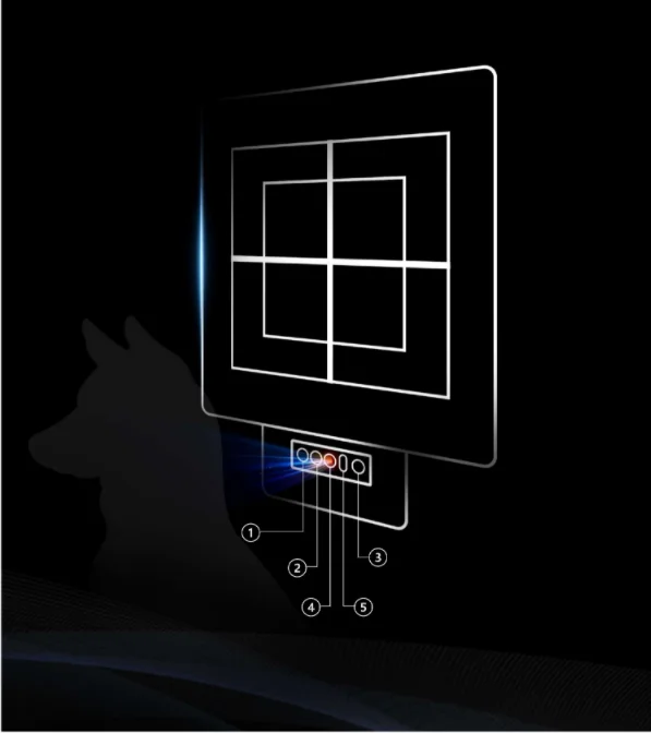
Al intelligent visual beam aligner
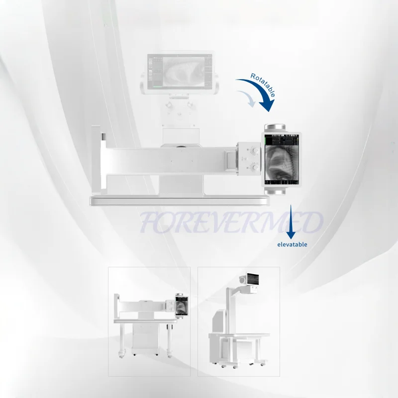
Rotary Arm
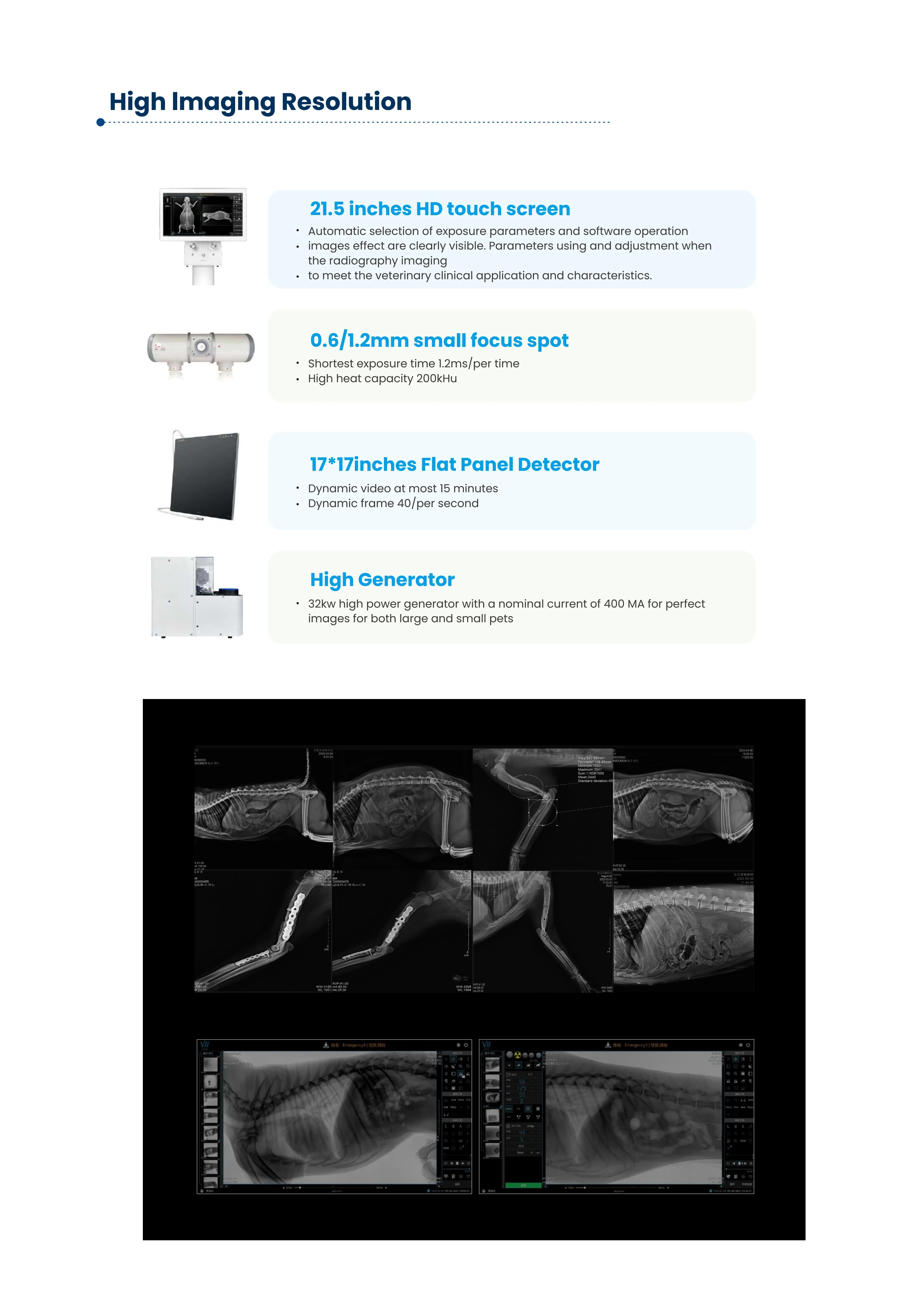
Multifunctionaland Dynamic videomore diagnosis information
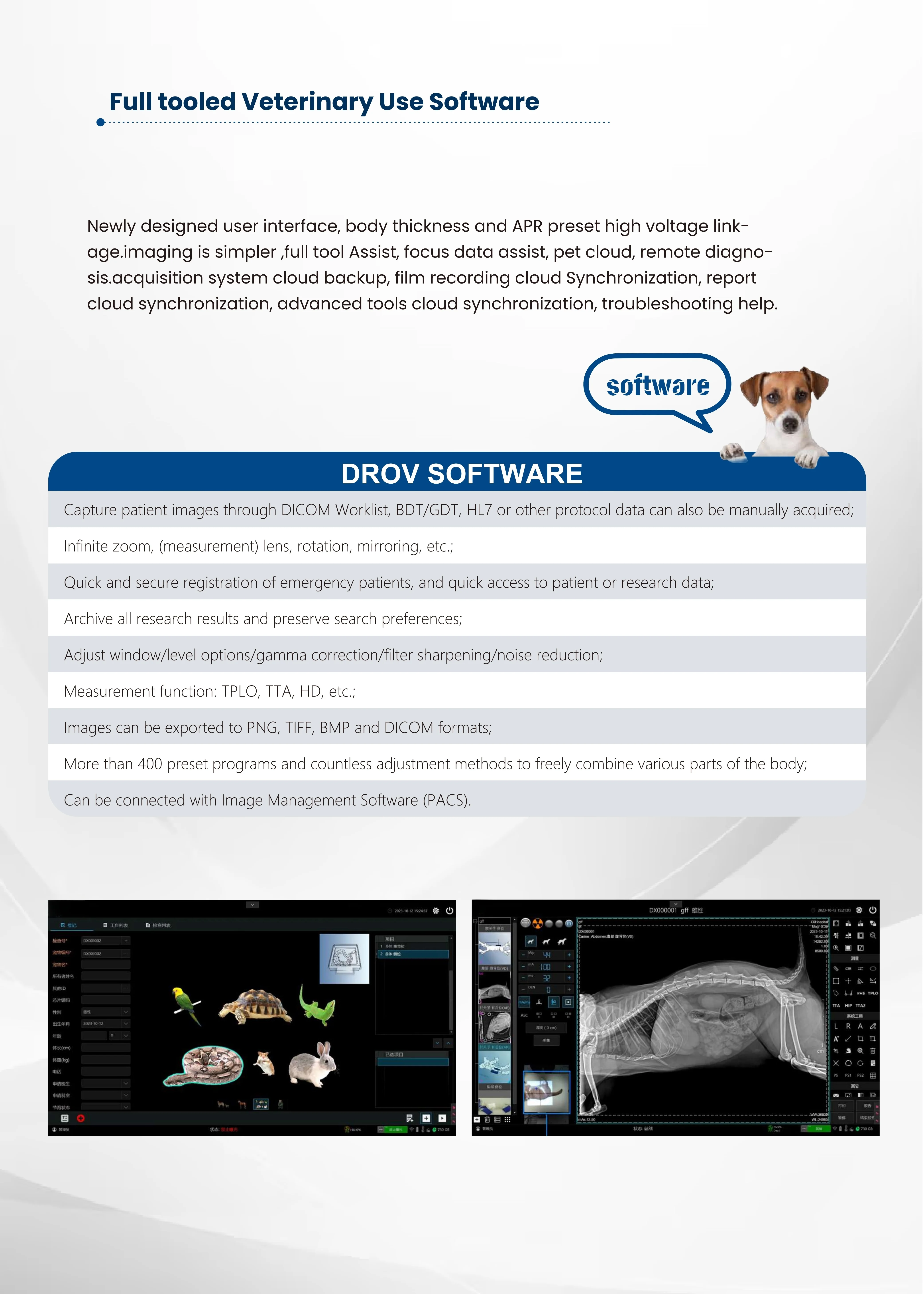
Detailed Pictures


RAQ
Q: 1. What kind of medical equipment does your company specialize in?
Q: 2. Which countries or regions do you export to?
Q: 3. What is your shipping method and delivery time?
Q: 4. What payment methods do you accept?
Q: 5. May we have a tour of your factory?
Q: 6. Do you provide after-sales service?
Q: 7. Can you provide customized solutions for medical devices?
Q: 8. What is your price range for medical equipment?
Q: 9. How can we guarantee the authenticity of your products?
Q: 10. What is your target market for medical devices?
Contact Information

Tel: +8615290808151

Phone: +8615290808151

Email: judy@yjcompany.com

Address: Wanda Center, Jinshui District, Zhengzhou, Henan, China

Whatsapp: +8615290808151

Skype: live:.cid.f4a1b4801c7fee8

Wechat: +8615290808151
Hot Tags: best veterinary digital radiography system, China, suppliers, manufacturers, factory, low price
|
No |
Item |
Quantity |
|
1 |
Veterinary use 32KW High Power Generator |
1 unit |
|
2 |
XRayTube |
1 unit |
|
3 |
Beam Aligner |
1 unit |
|
4 |
Metal Imaging Bed |
1 sheet |
|
5 |
Flat Panel Detector |
1 pair |
|
6 |
Desktop Computer set |
1 set |
|
7 |
15.6 touchable screen |
1 piece |
|
8 |
Vet Software |
1 set |
|
9 |
Lead Coat (Three-pieces) |
2 set |
|
10 |
Vet Position assistor |
2 pieces |
|
11 |
Cables and other accessories |
|
Product Parameters: |
||
|
No |
Parameters |
|
|
1 |
X Ray High Power Generator |
|
|
1.1 |
Power Supply Voltage: Single-phase 3-wire power cable 220V土22V |
|
|
1.2 |
Supply Frequency SOHz 土 l Hz |
|
|
1.3 |
Rated Power : 32Kw |
|
|
1.4 |
Max Output Current :400mA |
|
|
1.5 |
Operating frequency 300Hz |
|
|
1.6 |
Current regulation range :lOmA'""'--'400mA |
|
|
1.7 |
Tube voltage regulation range: (RAD)40-150kV, (Fluoro)40-110kV |
|
|
1.8 |
Exposure time range ; O.OOls'""'-'10.0s for imaging mode, 15minutes for dynamic mode |
|
|
1.9 |
Current time product range: O.lmAs-----320mAs |
|
|
2 |
Canon 72 40 X Ra y Tube |
|
|
2.1 |
Maximum peak voltage 125kv |
|
|
2.2 |
Spot Focus :Smallspot focus :0.6mm ,large spot focus :1.mm |
|
|
2.3 |
manode input power: 20kw for small sport focus/40kw for large spot focus |
|
|
2.4 |
Anode Speed : 2700rpm |
|
|
2.5 |
Target Angle 14° |
|
|
2.6 |
Anode heat capacity :200KHU |
|
|
3 |
Beam Al i g n e r |
|
|
3.1 |
Control method: Manual |
|
|
3.2 |
Radiation field Omm*Omm-430mm*430mm |
|
|
3.3 |
Power Supply :12V-24V |
|
|
3.4 |
light duration 30土ss |
|
|
3.5 |
inherentfiltration75kV: ?:1.0mmAI |
|
|
3.6 |
Weight: 6kg土0.5 |
|
|
4 |
Metal Imaging Bed (fixed straight arm +flo a t i n g t a b l e ) |
|
|
4.1 |
Table size : LXW 1510mmx635mm |
|
|
4.2 |
Total Height :1960mm |
|
|
4.3 |
Rotation angle: Perpendicular 90 to the right |
|
|
4.4 |
Floating Max size (Front to Behind):180mm Floating Maxsize (Left to Right) :240mm |
|
|
5 |
Iray 1717Z Flat Panel Detector |
|
|
5.1 |
Scintillator: Csl: TI |
|
|
5.2 |
Imaging region :430 x 430 mm2 |
|
|
5.3 |
Pixel array :3072 x 3072 |
|
|
5.4 |
Pixel size :139µm |
|
|
5.5 |
A/Dconversion : 16bits |
|
|
5.6 |
Energy range :50kV-11 OkV |
|
|
5.7 |
Date interface :GigE |
|
|
5.8 |
Trigger Mode :Syncin/Syncout |
|
|
5.9 |
Binning :1x1 /2x2/3x3 |
|
|
5.10 |
Sensitivity (ADC/nGy): > 15@PGA3 (1.2pC) Binning 3x3 > 0.85@PGA6{7.2pC)Binning lxl |
|
|
5.11 |
Dynamic range :>77dB@PGA6 1x1 |
|
|
5.12 |
MTF@RQAS: 55%@1.0LP/mm 30%@2.0LP/mm 14%@3.0LP/mm |
|
|
6 |
Veterinary Software |
|
|
6.1 |
Interface: 16languages supported ,work-list supported |
|
|
6.2 |
Capture patient images through DlCOM Worklist) BDT/GDT) HL7 or other protocol data can also be manually acquired; |
|
|
6.3 |
Infinite zoom,(measurement)lens, rotation, mirroring, etc.; |
|
|
6.4 |
Quick and secure registration of emergency patients, and quick access to patient or research data;, |
|
|
6.5 |
Archive all research results and preserve search preferences; |
|
|
6.6 |
Adjust window/level options/gamma correction/filter sharpening/noise reduction; |
|
|
6.7 |
Measurement function: TPLO, TTA, HD, etc.; |
|
|
6.8 |
lmages can be exported to PNG, TIFF, BMP and DlCOM formats |
|
|
6.9 |
More than 400 preset programs and countless adjustment methods to freely combine various parts of the body; |
|
|
6.10 |
Can be connected with Image Management Software (PACS). |
|
|
7 |
Computer set |
|
|
7.1 |
Intel Dual Core , WinlO operation system |
|
|
7.2 |
2T Hard Disk Drive |
|
|
7.3 |
24inches computer screen |
|
|
7.4 |
21.5 inches touchable screen in radiation room |
|


