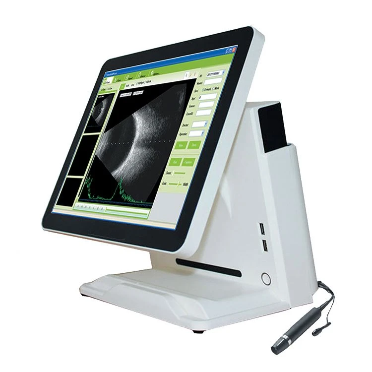
Ultrasound ophthalmology special diagnostic instrument
Ultrasound ophthalmic special diagnostic instrument is a special ophthalmic equipment used for intraocular disease diagnosis, measurement of ocular biological structure parameters and numerical calculation and design of intraocular lens, also known as ophthalmic AB ultrasound.
principle
The application of ophthalmic ultrasound in ophthalmic ultrasound diagnosis is to use the principle of ultrasonic ranging, and calculate the distance by measuring the time interval of the reflected waves at the interface of different tissues of the eyeball.
Usually can be divided into:
1. Axial length measurement: A ultrasonic probe of about 10MHz is usually used to identify the strong reflection of ultrasonic waves in the interface between the anterior chamber, lens, vitreous and retina, and measure the transmission time of ultrasonic waves in different tissues, and then according to the speed of sound in different tissues. Calculate the distance of each segment and obtain the axial length AL. The ophthalmic A-ultrasound probe is usually measured by the direct contact method or the water bath method. When the direct contact method is used, the contact part of the probe is the cornea (mucosal contact). When the water bath method is used, an eye cup is required.
2. Corneal thickness measurement: usually a 15MHz-20MHz corneal thickness probe is used to measure the time interval of the ultrasonic wave reflected at the front and back interfaces of the cornea, and then calculate the corneal thickness according to the sound speed of the cornea.
The basic principle of ophthalmic B-ultrasound imaging is the same as that of general-purpose B-mode ultrasound diagnostic equipment. According to its expected needs for the inspection of intraocular and orbital tissue structures, a high-frequency probe of 10MHz-25MHz is usually used, and its geometric size and acoustic window structure should be suitable for the structure of the human eye. The 0-13MHz ophthalmic B-ultrasound probe is used for imaging of intraocular and orbital tissues. 13-25MHz ophthalmic B-ultrasound probes are usually used to improve the scanning resolution of retinal tissue structures. The probe usually contacts the eyelid skin directly to image the eyeball and orbit.
Indications
1. Eye diseases: such as leukocysia, unclear refractive interstitium, retinal choroid detachment, fundus mass, eyeball atrophy, intraocular parasites, posterior scleritis, postoperative shallow anterior chamber, etc.
2. Orbital disease: such as proptosis, intraorbital tumor, inflammation and hemorrhage, eye movement disorder, etc.
3. Eye trauma: penetrating eyeball injury, eyeball rupture, intraocular hemorrhage, foreign body, etc.
4. Eye biometrics: such as axial measurement, intraocular lens power calculation, corneal thickness, etc.
5. Periorbital lesions, etc.