


Many friends will encounter such a situation when they go to the doctor to see the dentist: after the initial examination, the doctor will ask the patient to take x-rays. etc. These technical terms often confuse patients. Some patients even don't understand, why do I need to take a film when I can see a tooth and open my mouth? Is it necessary to take several pictures before and after a root canal treatment? In fact, as a routine auxiliary examination, dental film plays an irreplaceable role in the accurate diagnosis and treatment of doctors.
Filmmaking is irreplaceable
Our teeth are composed of a crown and a root. The crown is the part exposed in the mouth, while the lower root is buried in the jawbone like a tree root and is invisible to the naked eye. The outermost part of the crown is the enamel, which is the hardest tissue in the whole body, and together with the dentin on the inner side, it protects the core pulp tissue. Although the doctor can diagnose the lesions on the surface of the crown through intraoral examination, if the lesions develop into the inner dentin, pulp, or roots buried in the jaw, X-rays with perspective function are needed to observe. In addition to helping us understand the lesions of the teeth, X-ray films can also understand the "soil" around the teeth-the abnormalities in the alveolar bone. Therefore, X-ray film-assisted diagnosis can help doctors combine consultation and comprehensive analysis of clinical examination to improve the accuracy and success rate of diagnosis and treatment. Sometimes, x-rays are even the key to confirming the diagnosis or determining a treatment plan.
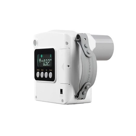
Dental tablets are roughly divided into three categories
The dental X-ray film that we use most often, doctors usually call it "small tooth film". a member of. When shooting, the unexposed film needs to be placed next to the teeth to be photographed in the patient's mouth. A perfect small dental film can show the images of the enamel, dentin, pulp and surrounding bone of the irradiated tooth, and can diagnose the lesions of the tooth and surrounding alveolar bone.
Panoramic film Mini small dental film can usually only see the range of 1~3 teeth. If you need to evaluate the condition of the whole mouth teeth and the surrounding jaw, you need to take a large film containing all the information of the teeth. This kind of X-ray film is similar to the panorama shooting function of a camera, and the teeth are seen at a glance by rotating around the jaw. Therefore, the information provided by panoramic films is very valuable in the presence of multiple tooth lesions, bone resorption in periodontal disease, treatment planning for restorative orthodontics, surgical wisdom tooth extraction, and mandibular lesions.
Dental CT Dental CT is a new star in oral imaging diagnostic examination in recent years, especially suitable for three-dimensional imaging of dental hard tissues. Since having it, dentists can finally see three-dimensional teeth without pulling them out. Dental CT provides very valuable spatial information for the complex overlapping structures that are unclear in the above two 2D images. Especially for the evaluation of complex and difficult root canal systems, impacted teeth, implant sites, etc., dental CT examinations are becoming more and more common.

Radiation dose is very low
Root canal treatment If caries is not treated early, root canal treatment is required when the dental pulp develops to cause inflammation. Before and after root canal treatment, 3 to 5 small dental films need to be taken. The preoperative film is used to judge the progress of the disease and whether the bone below the root is involved; the intraoperative tracer film is used to judge the preparation of the root canal and whether the entire root canal has been successfully opened; the postoperative film is used to judge the filling of the root canal Is it perfect. Sometimes it is necessary to take additional follow-up films several months after the operation to observe the healing situation. Many patients complain about taking too many films. In fact, doctors use X-ray examinations to escort patients throughout the treatment process and improve the success rate of treatment. For some difficult root canals, such as multiple canal systems, curved root canals, and C-shaped root canals, dental CT is required.
Wisdom tooth extraction Many young friends have encountered wisdom tooth inflammation. The suggestion of dentists is to pull out wisdom teeth early and easily. But before wisdom teeth are extracted, X-ray examination is essential, which can let us know how deep the wisdom teeth are buried, how many roots there are, whether there will be any resistance before extraction, and the distance from the nerve canal in the jawbone. How close, will it be easy to accidentally injure and so on. For some complicated wisdom teeth, dental CT will tell us more three-dimensional information, such as where to dig a hole to find it nearest, such as which direction the neural tube is out of shape in the wisdom teeth, how to avoid it, and so on.
Dental CT before and after implant surgery has promoted the development of dental implants. Now we can judge not only the height of the bone but also the thickness of the bone, and we can also evaluate the conditions around the "soil" of the implant, making the implant safer and safer. After the implant is finished, take a post-operative photo to see if the implant is in the desired position.
Finally, it should be emphasized that the radiation dose of dental X-ray film examination is very low compared with that of general examination, so patients and friends do not need to worry. X-rays are often necessary when doctors feel they need them for diagnosis and treatment planning.


