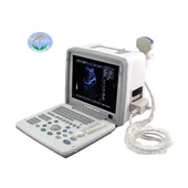
What is a panoramic dental X-ray machine
When visiting a dentist, you will usually be X-rayed, a piece of plastic is placed in your mouth and bitten with your teeth, and a few pictures of one or more of your teeth are taken. A dentist will usually do several such X-rays to identify conditions that may affect different parts of your mouth.
In contrast to this traditional radiograph, a dental panoramic X-ray creates a single image of the entire oral cavity: the upper or lower jaw, the temporomandibular joint, the entire set of teeth, the nasal area and the sinuses. This kind of photo reflects the curved structure of the jaw flatly, making it easier to analyze each part.
Why use panoramic X-ray?
Because panoramic X-rays can show the entire oral cavity in one picture, there is no need to generate detailed pictures showing caries. Such X-ray pictures can show bone abnormalities and fractures, cysts, impacted teeth, infections and tumors. A dentist who suspects any such problems can take a panoramic X-ray of the patient. Dentists may also use this method of imaging in the following situations: planning to adopt braces, implant dental fillings, and wear dentures.
How to take this X-ray photo
Different from traditional intraoral X-ray photography, dental panoramic X-ray is an extraoral photography, which means that the imaging machine and film are outside the oral cavity. According to the Radiological Society of North America (RSNA), a dental panoramic X-ray machine projects light through your mouth onto a film or detector that rotates so as to face the X-ray tube.
The basic design of the dental panoramic X-ray machine is that the picture tube is mounted on a horizontal rod that can point to one side of the patient's cheek, and the opposite horizontal rod points to the other side containing the X-ray film or detector. Generally speaking, the head is positioned by the lower handle, forehead and side, and a pair of swallowing stops is used to keep the mouth open. Then, the rod of the X-ray machine will rotate in a semicircle around the patient's head, starting from one side of the lower handle and ending on the other side.
Benefits and risks of X-ray
Panoramic X-ray allows the dentist to view the patient’s entire oral cavity on a single photo in a relatively short time. However, according to the Society of Pediatric Radiology, the radiation exposure of one panoramic X-ray is 0.02 microsieverts, which is four times the 0.005 microsieverts produced by four bite-film X-rays taken during regular inspections. If you are concerned about radiation exposure, please inform your dentist and ask if other shooting methods can be used.