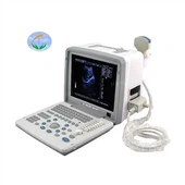
Three-dimensional color Doppler ultrasound is a kind of color Doppler ultrasound, which is a three-dimensional dynamic display. The superiority of three-dimensional color Doppler ultrasound is mainly reflected in the sensitivity and high display rate of uterine artery and ovarian blood flow. ② Shorten the inspection time and obtain accurate Doppler spectrum. ③ No need to fill the bladder. ④Do not interfere with obesity, abdominal scars, and intestinal cavity inflation. ⑤ Use the activity of the probe tip to find the tender parts of the pelvic organs to determine whether there are adhesions in the pelvic cavity.
Three-dimensional color Doppler ultrasound surface imaging is used in obstetric examinations. It can not only observe the growth process of the fetus, but also check the changes of the placenta, amniotic fluid and umbilical cord. More importantly, it can be used as the main means of diagnosing fetal malformations. Due to the large contrast between the tissue structure and the liquid gray scale, the three-dimensional shape, surface characteristics, and spatial position relationship of the suspicious structure can be clearly displayed, and the three-dimensional image of the fetus in the uterus can be provided. Three-dimensional reconstruction includes surface imaging, transparent imaging and multi-plane imaging modes.
Features of three-dimensional color Doppler ultrasound
(1) Adding coronal section images on the basis of retaining two-dimensional imaging.
(2) Stereo positioning, axis adjustment, ABC three axis planes can be adjusted at will until the best image is displayed.
(3) Stereoscopic imaging, dynamic and intuitive; real-time dynamic observation of the fetal head, body surface and internal organs, the image is clear, accurate and reliable.
(4) The cutting function can keep the key points of the image; cut off the useless parts, and perform three-dimensional reconstruction and display of the suspicious parts.
(5) Rotation function, multi-faceted observation; 360° rotation function for front and back, left and right, up and down, and comprehensive observation of images in different directions.
(6) It can take exquisite photos of the fetus, record expression changes and burn a disc as data for preservation, and keep it as a permanent memorial.
(7) It can show the three-dimensional relationship and adjacent relationship of different levels of lesions. The application of three-dimensional color Doppler ultrasound in obstetrics provides a wealth of image information for clinical ultrasound diagnosis. The fetus is surrounded by liquid in the amniotic cavity is a good imaging condition for three-dimensional ultrasound. The image is three-dimensional and intuitive, and the angle can be adjusted arbitrarily. Suspicious structures can be observed by rotating, and the malformations of the general structure of the fetus can be seen at a glance, which greatly improves the quality of diagnosis and treatment, and reduces misdiagnosis or missed diagnosis.
3D color Doppler ultrasound time
Generally, it is an ideal time to do three-dimensional color Doppler ultrasound to check the development of the fetus to eliminate abnormalities from 22 to 26 weeks of pregnancy.